Cytology

Return to Search Page

images/actinocyte.JPG
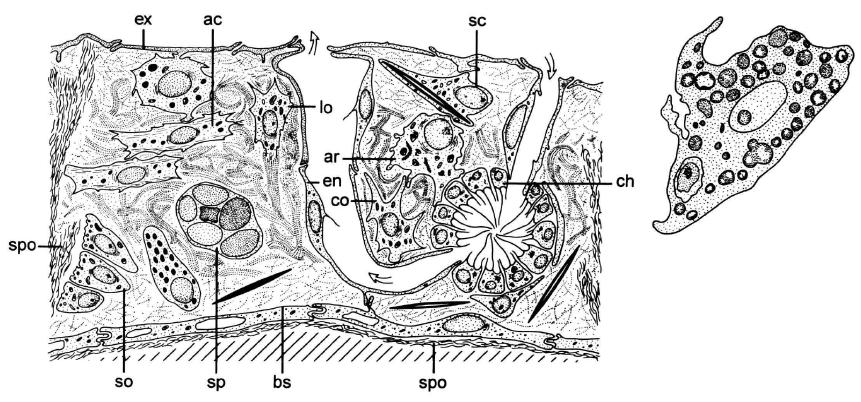
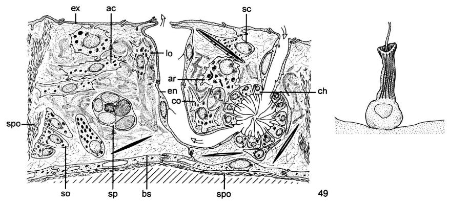
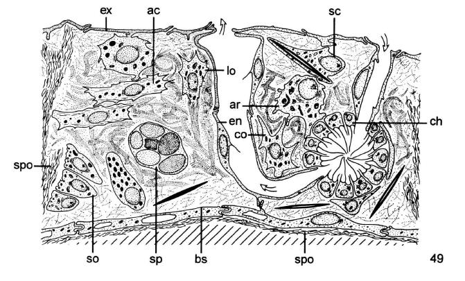
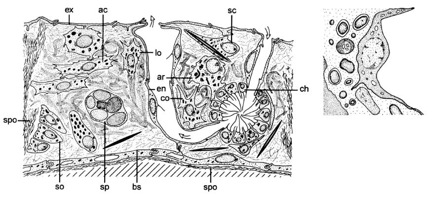
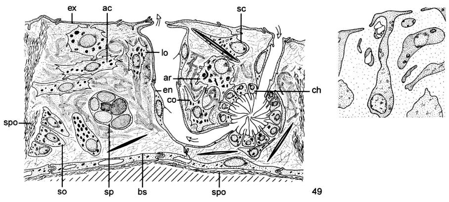
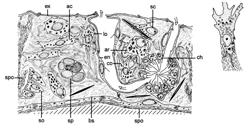
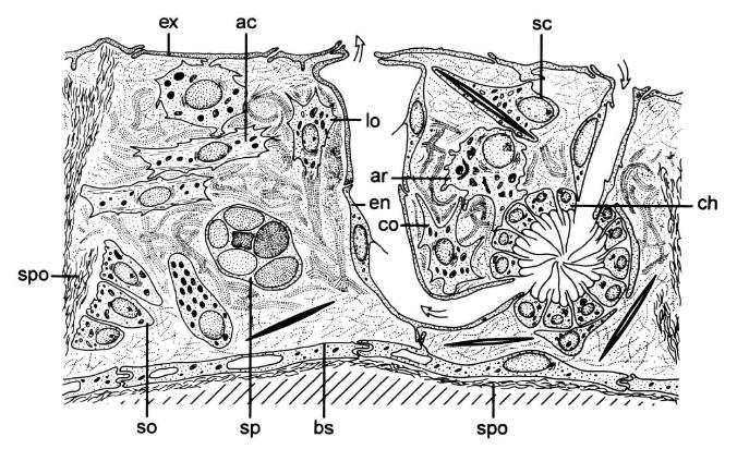
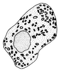
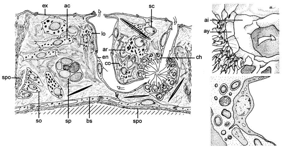
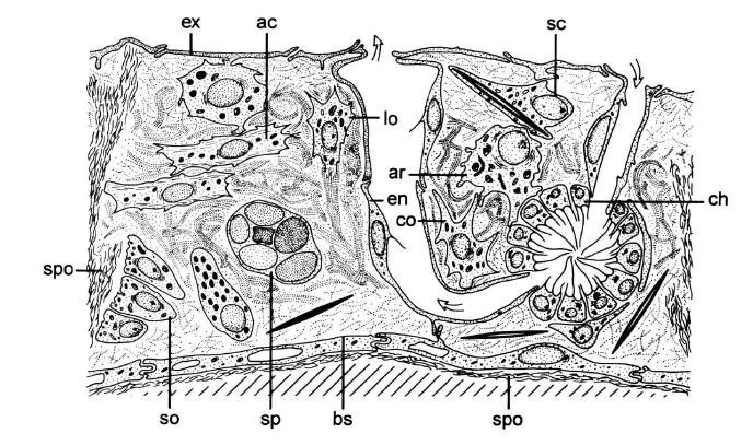
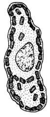
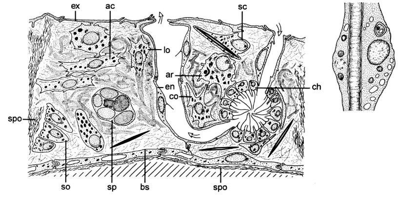
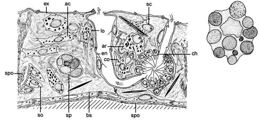
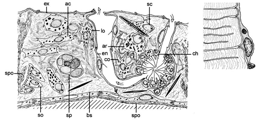
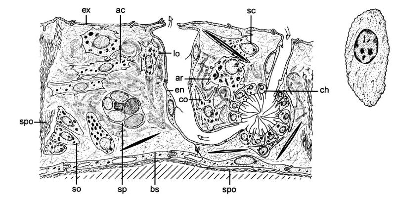
Word:
Actinocyte

Description:
Elongated contractile cell often grouped in sphincter- like structure around the osculum, below the sieve- plates, and around the large exhalant canals. Characterized by numerous filaments of actin within their cytoplasm. New term for the contractile cell (ac in figure).

Synonyms:
Myocyte

Category:
Cytology


images/apopinacocyte.JPGWord:
Apopinacocyte

Description:
Endopinacocyte lining the exhalant canal (ai in figure).

Category:
Cytology


No image available.

Word:
Apopinacoderm

Description:
Surface lined by apopinacocytes.

Category:
Cytology


images/apopylar cell.JPGWord:
Apopylar cell

Description:
Cell surrounding the apopyle and located between the choanocytes and the apopinacocytes (ay in figure).

Category:
Cytology


images/archaeocyte.JPGWord:
Archaeocyte

Description:
Amoeboid cell with large nucleolus and capable of phagocytosis. May differentiate into other types of cells (ar in figure).

Category:
Cytology


images/bacteriocyte.JPGWord:
Bacteriocyte

Description:
Cell containing prokaryotic microsymbionts

Category:
Cytology


images/basopinacocyte.JPGWord:
Basopinacocyte

Description:
Pinacocyte affixing the sponge to the substratum by external secretion of a collagenous matrix (bs in figure).

Category:
Cytology


images/central cell.JPGWord:
Central cell

Description:
Single cell located at the apopyle of choanocyte chambers

Category:
Cytology


images/choanoblast.JPGWord:
Choanoblast

Description:
Cell that buds anucleate outgrowth called COLLAR BODY; in Hexactinellida

Category:
Cytology


images/choanocyte.JPGWord:
Choanocyte

Description:
Cell having a flagellum that is surrounded by a collar of cytoplasmic microvilli linked by bridges of glycocalyx. Several are typically arranged in spherical, ovoid, or elongated chambers. Fundamental cell of sponges (ch in figure).

Category:
Cytology


No image available.

Word:
Choanoderm

Description:
Surface lined by choanocytes.

Category:
Cytology


images/collar body.JPGWord:
Collar body

Description:
Anucleate choanocyte in hexactinellids.

Category:
Cytology


images/collencyte.JPGWord:
Collencyte

Description:
Cell with branching pseudopods, involved in the secretion of collagen (co in figure).

Category:
Cytology


images/cystencyte.JPGWord:
Cystencyte

Description:
Cell with a single large vesicle containing amorphous material identified as polysaccharide and occupying most of the cell volume

Category:
Cytology


images/endopinacocyte.JPGWord:
Endopinacocyte

Description:
Pinacocyte lining the inhalant and exhalant canals. In some Demospongiae orders, it has a flagellum (en in figure).

Category:
Cytology


No image available.

Word:
Endopinacoderm

Description:
Surface lined by endopinacocytes.

Category:
Cytology


No image available.

Word:
Eosinophilic cell

Description:
Cell with inclusions that react positively with eosine.

Category:
Cytology


images/exopinacocyte.JPGWord:
Exopinacocyte

Description:
Fusiform or T-shaped pinacocyte covering the free surface of a sponge. In exceptional cases, it has a flagellum (Homoscleromorpha) (ex in figure).

Category:
Cytology


No image available.

Word:
Fuchsinophilic cell

Description:
Cell with inclusions that react positively with acid fuchsin.

Category:
Cytology


images/globoferous cell.JPGWord:
Globoferous cell

Description:
Cell with a single large globule, a reduced cytoplasm, and a small conical nucleus (Clathriidae)

Category:
Cytology


images/glycocyte.JPGWord:
Glycocyte

Description:
Cell with conspicuous dictyosomes, characterized by the presence of glycogen rosettes and osmiophilic inclusions. Also known as gray cell

Synonyms:
Gray cell

Category:
Cytology


images/lophocyte.JPGWord:
Lophocyte

Description:
Collencyte with a characteristic tuft of collagen fibrils attached to the posterior pole (lo in figure).

Category:
Cytology


images/mesohyl.JPGWord:
Mesohyl

Description:
Part of sponge enclosed by pinacoderm and choanoderm (= "mesenchyme," term to be rejected for sponges).

Category:
Cytology


No image available.

Word:
Mesolamella

Description:
Sheet of collagen separating the choanochambers of hexactinellids.

Category:
Cytology


images/microgranular cell.JPGWord:
Microgranular cell

Description:
A cell with cytoplasm filled with small dense granules

Category:
Cytology


images/microsymbiont.JPGWord:
Microsymbiont

Description:
Prokaryotic or more rarely eukaryotic microorganism living inside a sponge. May be unicellular or multicellular filamentous, with or without pigments, and may occur intra- or intercellularly

Category:
Cytology


images/pinacocyte.JPGWord:
Pinacocyte

Description:
Cell delimiting the sponge from the external milieu and always only in a layer one-cell deep

Category:
Cytology


images/pinacoderm.JPGWord:
Pinacoderm

Description:
Surface lined by pinacocytes (ex and en in figure).

Category:
Cytology


images/pocket cell.JPGWord:
Pocket cell

Description:
Special kind of bacteriocyte that surrounds an area of the intercellular matrix, including microsymbionts

Category:
Cytology


images/porocyte.JPGWord:
Porocyte

Description:
Cell surrounding a pore (ostium)

Category:
Cytology


No image available.

Word:
Prosopinacocyte

Description:
Endopinacocyte lining the inhalant canals.

Category:
Cytology


No image available.

Word:
Prosopinacoderm

Description:
Surface lined by prosopinacocytes.

Category:
Cytology


images/rhabdiferous cell.JPGWord:
Rhabdiferous cell

Description:
Large cell with numerous rod-like inclu sions parallel to the long axis of the cell containing acid mucopolysaccharides

Category:
Cytology


images/sclerocyte.JPGWord:
Sclerocyte

Description:
Cell involved in spicule secretion. In Demospon- ges, with intracellular secretion, sclerocytes are characterized by numerous mitochondria and the presence of spicule-axial filaments. In Calcarea, where secretion is extracellular, sclerocytes have septate junctions between them (sc in figure).

Category:
Cytology


images/spherulous cell.JPGWord:
Spherulous cell

Description:
Cell filled with large round spherules that occupy almost the entire cell body, compressing the cytoplasm into thin sheets (sp in figure).

Category:
Cytology


images/spongocyte.JPGWord:
Spongocyte

Description:
Cell secreting spongin fibers (so (spo = spongin) in figure).

Category:
Cytology


images/thesocyte.JPGWord:
Thesocyte

Description:
Vitelline-rich archaeocyte in a sponge gemmule (th in figure).

Category:
Cytology


No image available.

Word:
Trabecular tissue

Description:
Syncytia in hexactinellids occurring around the choanochambers.

Category:
Cytology


images/trophocyte.JPGWord:
Trophocyte

Description:
Nurse cell involved in the initial stages of gemmule and oocyte formation.

Category:
Cytology

Return to Search Page